Dental imaging can provide important information that may not be visible during a routine oral examination. One commonly used examination is an orthopantomogram, usually called an OPG or panoramic dental X-ray. It creates a broad two-dimensional image of the teeth, upper and lower jaws, and surrounding structures in a single scan.
For patients who have been advised to undergo an OPG dental X-ray Dubai examination, understanding what the scan involves, why it may be recommended, and how to prepare can make the appointment easier.
What Is an OPG Dental X-ray?
An OPG is a panoramic dental X-ray that captures a wide view of the mouth and jaw in one image.
Unlike conventional intraoral dental X-rays, where the detector is positioned inside the mouth, an OPG uses a machine that rotates around the patient’s head while capturing the required images.
The resulting image can show the upper and lower teeth, jawbones, and certain surrounding structures. Because it covers a relatively large area, an OPG can provide dentists with a useful overall view of the mouth.
Why Is an OPG Recommended?
Dentists and oral surgeons may recommend panoramic imaging for several reasons.
An OPG can be useful when evaluating impacted teeth, including wisdom teeth, jaw abnormalities, certain cysts, advanced periodontal disease, and other dental or oral conditions. It may also be used when planning treatments such as braces, dentures, tooth extractions, or dental implants.
The need for an X-ray should depend on the patient’s individual dental condition and the information required for diagnosis or treatment planning. Current American Dental Association recommendations emphasize that dental X-rays should be ordered when clinically necessary rather than according to a routine schedule that applies to everyone.
What Can an OPG Show?
An OPG provides a broad overview rather than highly detailed images of individual teeth.
Depending on the quality of the image and the clinical situation, it may help a dentist assess:
- Tooth position and development
- Impacted wisdom teeth
- Jawbone structure
- Certain cysts or lesions
- Advanced periodontal changes
- Some jaw abnormalities
- The general position of teeth before orthodontic treatment
- Structures relevant to extraction or implant planning
An OPG may also provide information about parts of the maxillary sinuses and other surrounding structures.
However, an OPG does not provide the same level of detail as every other dental imaging method. If a dentist needs more precise information about a particular tooth or area, additional intraoral X-rays or three-dimensional imaging such as cone-beam CT may be considered.
How Does an OPG Work?
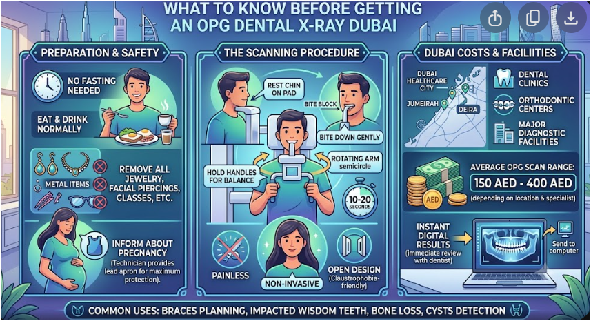
During an OPG dental X-ray Dubai appointment, you will usually stand or sit in the center of the panoramic X-ray machine.
The technician carefully positions your head using supports to keep it correctly aligned.
You may be asked to bite gently on a positioning device to help maintain the correct relationship between your upper and lower teeth.
The X-ray tube and detector then move around your head in a semicircular path while the image is captured. The actual image acquisition is typically very quick, often lasting only several seconds.
Remaining still during the scan is important because movement can affect image quality.
Is an OPG X-ray Painful?
An OPG examination is generally painless and non-invasive.
You may need to keep your head and body in a particular position for a short period, but the scan itself does not normally cause discomfort.
For patients who have a sensitive gag reflex, panoramic imaging can sometimes be more comfortable than conventional intraoral X-rays because the detector does not need to be placed inside the mouth.
If you have difficulty standing for the examination, tell the imaging professional. Some panoramic systems can accommodate patients who need to be seated.
How Should You Prepare?
An OPG generally requires little or no special preparation.
Before the examination, you may be asked to remove jewelry, glasses, or other metal objects around the head and neck that could interfere with the image.
You can normally eat and drink as usual unless your dentist or imaging provider gives you different instructions.
It is also useful to bring previous dental X-rays or reports when available, particularly if your dentist wants to compare earlier images.
What About Radiation Exposure?
Because an OPG is an X-ray examination, it uses a small amount of ionizing radiation.
The radiation dose from panoramic dental imaging is relatively low. RadiologyInfo lists an approximate effective dose of 0.025 mSv for a panoramic X-ray, although the actual dose can vary depending on equipment and imaging technique.
Dental professionals are encouraged to use X-rays only when clinically necessary and to keep radiation exposure as low as reasonably achievable while obtaining diagnostically useful images.
If you are pregnant or think you may be pregnant, inform your dentist or imaging professional before the examination.
What Happens After the OPG?
Once the image has been captured, it is reviewed by a qualified dental or radiology professional.
The dentist may use the image to assess teeth and jaw structures, plan treatment, or determine whether additional imaging is needed.
An OPG may sometimes reveal an abnormality that requires closer examination. This does not automatically mean that a serious condition is present.
If a more detailed assessment is required, your dentist may recommend another type of dental X-ray, CBCT, CT, MRI, or another appropriate examination.
Choosing an OPG Dental X-ray Dubai Provider
When selecting an OPG dental X-ray Dubai provider, consider whether the facility has appropriate panoramic imaging equipment, trained professionals, suitable patient-positioning procedures, and a clear process for providing images and reports.
It is also helpful to confirm whether the facility can perform additional dental imaging if your dentist believes it may be necessary.
For children or patients with special positioning requirements, ask whether the imaging team can accommodate their individual needs.
OPG vs. Other Dental X-rays
An OPG provides a broad overview of the teeth and jaws, while intraoral X-rays can provide more detailed images of individual teeth or specific areas.
CBCT, meanwhile, produces three-dimensional images and may be selected when detailed information about dental structures or jaw anatomy is required for certain procedures.
The appropriate imaging method depends on the clinical question. One type of dental X-ray does not replace every other type.
When Should You Have an OPG?
An OPG should generally be performed when recommended by a dentist or other qualified healthcare professional.
It may be useful before certain dental treatments or when a dentist needs a broad overview of the teeth and jaw.
However, routine X-rays are not necessary at the same frequency for every patient. Oral health, age, symptoms, disease risk, previous findings, and treatment needs can all influence the decision.
Making an Informed Decision
An OPG dental X-ray Dubai examination provides a broad view of the teeth and jaw in a single image and can support dental diagnosis and treatment planning.
The examination is quick, non-invasive, and generally painless, with relatively low radiation exposure.
Before your appointment, follow any instructions provided by your dentist, remove objects that could interfere with imaging, and inform the imaging team if you may be pregnant.
Most importantly, dental X-rays should be performed for an appropriate clinical reason. When the examination is properly selected, performed, and interpreted, an OPG can provide useful information that helps dentists evaluate oral structures and plan appropriate care.
Share this content:
Post Comment